
Mitochondria Imaging Services
Mitochondria is a kind of organelle with double-layer membrane structure, which is called "cell power chamber". Mitochondrial morphology is an active regulation and dynamic feature through mitochondrial dynamic changes, including mitochondrial autophagy, mitochondrial fission, fusion and so on. The changes of mitochondrial morphology are related to calcium homeostasis, lipid modification, regulation of oxidative metabolism and cell death. Therefore, it is very important to analyze the changes of mitochondrial shape.
 Figure 1. Section of mitochondria.
Mitochondria Morphology Imaging Analysis
Analyzing the structure of mitochondria is a decisive factor in describing their state and function. Usually, different fluorescent dyes can be used to stain them, and then the mitochondrial morphology can be observed in living cells through a microscope. Initially, mitochondrial morphology analysis used manual classification and evaluation methods or automatic methods with limited types of features to analyze still images. With the development of mitochondrial imaging technology and fluorescent dyes, the qualitative observation of mitochondrial morphology has been greatly improved. In recent years, live-cell imaging of mitochondria has shown that these organelles are highly dynamic, which poses a serious challenge for high-resolution imaging of mitochondria. This problem was not solved until the advent of confocal microscopes with high-speed acquisition speeds.
CD BioSciences can synthesize a variety of fluorescent dyes for you according to your needs, and use a confocal microscope to perform steady-state and time-lapse imaging of mitochondria in living cells, and then we use ImageJ software to automatically analyze the images and generate data analysis reports.
Mitochondria Morphology Imaging Analysis Workflow
The following are the specific experimental steps of mitochondria morphology imaging analysis:

Synthetic fluorescent dyes
We will design and synthesize a variety of fluorescent dyes for you according to your needs, and characterize them by NMR, dynamic light scattering and GC.
Step 1
Cell culture
The target cells were cultured in DMEM-HG medium under standard conditions, supplemented with fetal bovine serum and alanyl-glutamine. After the cells reached 80% confluence, the cells were washed with PBS and replaced with DMEM containing glucose but not phenol red.
Step 2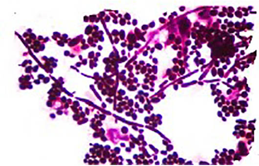
Live cell staining
After determining the minimum dye concentration required to obtain a high signal-to-noise ratio image, the cells were stained with a fluorescent dye in phenol red-free DMEM for 30 minutes at 37 °C and then replaced with a new medium.
Step 3
Confocal fluorescence imaging
The stained cells were seeded into a 96-well plate and imaged by a confocal microscope to monitor the morphological changes of mitochondria during the cell event and obtain images at different time points.
Step 4
Image analysis
We use image analysis software to preprocess and segment the collected images, then perform mitochondrial morphology measurement and calculation, and generate readable reports according to your needs.
Step 5Delivery
Number of mitochondria per cell
Mitochondrial mass
Mitochondrial shape factor
Mitochondrial size
Mitochondrial aspect ratio
Provide other data according to your needs
Our Advantages
- Capable of large-scale mitochondrial morphology analysis
- In-depth analysis of mitochondrial characteristics of any cell type
- Ability to capture live high-resolution images over time
- Experienced scientists provide experimental consultation
- Reasonable price and short turnaround time
CD BioSciences has a professional team and advanced imaging equipment. The entire process of mitochondria morphology imaging analysis is operated by experienced technicians to ensure the accuracy of the experiment. If you have any needs, please feel free to contact us. We will provide you with high-quality services that meet your needs.
- Bosch, Anna, and Maria Calvo. "Automated quantitative analysis of mitochondrial morphology." Computer Optimized Microscopy. Humana, New York, NY, 2019. 99-115.
- Leonard, Anthony P., et al. "Quantitative analysis of mitochondrial morphology and membrane potential in living cells using high-content imaging, machine learning, and morphological binning." Biochimica et Biophysica Acta (BBA)-Molecular Cell Research 1853.2 (2015): 348-360.
- Mitra, Kasturi, and Jennifer Lippincott‐Schwartz. "Analysis of mitochondrial dynamics and functions using imaging approaches." Current protocols in cell biology 46.1 (2010): 4-25.
*If your organization requires the signing of a confidentiality agreement, please contact us by email.
Please note: Our services can only be used for research purposes. Do not use in diagnostic or therapeutic procedures!