
Antibody Internalization Imaging Services
Antibody internalization is an important technique for studying the transport and distribution of antibodies inside cells. Antibodies are used as therapeutic agents that can target specific sites or cells to effectively treat a wide range of diseases, including autoimmune diseases, cancer, inflammation, cardiovascular diseases, and infectious diseases. Antibody internalization imaging techniques observe and quantify antibody transport and distribution inside cells in real time by labeling antibodies or receptors. These labeling techniques allow researchers to follow the internalization process of antibodies and study their kinetic properties, internalization rates and pathways, and status in different cell types and disease states.
 Fig. 1 Internalization interplay between two anti-EphA2 mAbs monitored by immunofluorescence (Liao-Chan S, et al. 2015).
Antibody Internalization Imaging Services
CD BioSciences provides antibody internalization imaging services to rapidly quantify the antibody internalization process. We utilize real-time live cell analysis technology for antibody screening and functional characterization to provide you with accurate and reliable data.
Screening of ADC drug candidates
Understanding the internalization capacity of antibodies and comparing the uptake of different antibodies are key metrics for early screening and optimization of ADC drugs. Our antibody internalization imaging service can help clients screen ADC drug candidates.
- Provide clients with a diverse selection of cell models, including tumor cell lines and modified cell lines.
- Provide a variety of assay platforms such as flow cytometry, confocal microscopy and high internalization imaging to meet different experimental needs.
- Our antibody internalization imaging service has been validated on multiple targets.
Dose-Response Testing
CD BioSciences' antibody internalization assay based on live cell imaging allows for quantitative dose-response testing
- Visualize and detect changes in antibody internalization over time at different concentrations.
- Compare the degree of internalization at different antibody concentrations, quantitatively assess the internalization efficiency of the antibody, and establish a concentration-internalization curve.
Pharmacological dynamic quantitative analysis
- Using a simple mix-and-read protocol.
- Evaluate time-dependent changes in antibody internalization by treating cells with increasing concentrations of antibody.
- Construct concentration-dependent curves.
- Provide data for quantitative analysis of pharmacological dynamics.

Antibody Internalization Imaging Workflow
CD BioSciences' antibody internalization imaging utilizes the principle of antibody internalization, labeling fluorescent dyes with pH-sensitive antibodies, and then using live cell imaging instruments to monitor the dynamics of antibody internalization in real time in a 96-well plate.

Seed cells into 96-well or 384-well plate and leave to adhere.
Step 1
Label test antibody and incubate to allow conjugation.
Step 2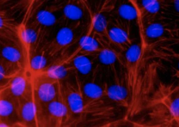
Add antibody mix to cell plate.
Step 3
Live cell fluorescent imaging and analysis.
Step 4Deliverables
- Antibody internalization imaging raw data
- Pharmacokinetic data report
- Processed data, graphs and summary reports
Our Advantages
- Unlike endpoint assays, a single experiment simultaneously evaluates time-dependent changes in antibody internalization and concentration-dependent curves, allowing for dynamic quantitative analysis in pharmacology.
- Real-time observation of live cells, continuous monitoring up to 72 hours, minimum detection interval of 15 minutes.
- Can be used for high throughput screening of antibodies.
CD BioSciences enables real-time, quantitative observation of antibody internalization behavior and obtains important information about intracellular transport and translocation mechanisms. If you have any needs, please feel free to contact us. We can also customize brain imaging services for you according to specific needs.
- Liao-Chan S, et al. Quantitative assessment of antibody internalization with novel monoclonal antibodies against Alexa fluorophores. PLoS One, 2015, 10(4):e0124708.
*If your organization requires the signing of a confidentiality agreement, please contact us by email.
Please note: Our services can only be used for research purposes. Do not use in diagnostic or therapeutic procedures!