
Angiogenesis Imaging Services
Angiogenesis is a physiological process, which is an essential part of many normal (reproductive and wound healing) and pathological processes (diabetic retinopathy, rheumatoid arthritis, tumor growth and metastasis). The effect of angiogenesis inhibitor on cell differentiation can be evaluated by matrix gel differentiation test. Matrigel differentiation test is an environment for simulating the process of natural angiogenesis in vitro. In vitro angiogenesis analysis provides a platform for evaluating the effects of Pro angiogenic compounds or anti-angiogenic compounds.
 Figure 1. The growth of blood vessels in cancerous tissue or cells is also known as angiogenesis.
Angiogenesis Imaging Analysis
Tube formation analysis is a widely used in vitro tool for obtaining angiogenesis in a simple, cost-effective and repeatable way. CD BioSciences' high-content imaging technology allows the visualization of angiogenesis structure, 3D reconstruction and complex analysis of angiogenesis and new vessel germination, which can be used for the comparative study of disease phenotypes and composite effects.
Angiogenesis Imaging Analysis Workflow
Angiogenesis
Endothelial cells (primary, cell line, or iPSC-derived) are seeded in the microfluidic top perfusion channel and attached to the ECM gel. As the cells proliferate, they form endothelial capillaries.
Add Inducing Compound
After the blood vessel is formed, the pro-angiogenic factor mixture is added to the bottom perfusion channel, located on the other side of the endothelial blood vessel. The resulting gradient of angiogenic compounds leads to the induction of angiogenic buds.
Imaging
Vessel cells and buds were fixed with 4% formaldehyde and stained with corresponding antibodies, and then the cells were imaged using a high-content imaging system. The nucleus was imaged with the DAPI channel, and the angiogenic buds were imaged with the FITC channel, with exposure at 100 milliseconds and 400 milliseconds, respectively.
Image Analysis
Use the custom module editor in the high-content image acquisition and analysis software to analyze the images and generate readable analysis reports.

Angiogenesis
Step 1
Add inducing compound
Step 2
Imaging
Step 3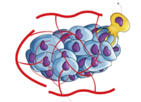
Image analysis
Step 4Delivery
Image of angiogenic bud
Angiogenic sprout volume
Number of angiogenic buds
Other relevant data
Our Advantages
- Visualize and quantitatively analyze angiogenic buds
- Generate high-throughput image data
- Experienced scientists provide experimental consultation
- Reasonable price and short turnaround time
CD BioSciences has a professional team and advanced imaging equipment. The entire process of angiogenesis imaging analysis is operated by experienced technicians to ensure the accuracy of the experiment. If you have any needs, please feel free to contact us. We can provide you with high-quality angiogenesis imaging analysis services, making it a quantitative tool for angiogenesis in the fields of embryonic development, cancer, wound healing and tissue repair.
- Valavanis I, Goudas T, Michailidou M, et al. A Novel Image Analysis Methodology for the Evaluation of Angiogenesis in Matrigel Assays and Screening of Angiogenesis-Modulating Compounds[C]//IFIP International Conference on Artificial Intelligence Applications and Innovations. Springer, Cham, 2015: 61-71.
- Boizeau M L, Fons P, Cousseins L, et al. Automated image analysis of in vitro angiogenesis assay[J]. Journal of laboratory automation, 2013, 18(5): 411-415.
*If your organization requires the signing of a confidentiality agreement, please contact us by email.
Please note: Our services can only be used for research purposes. Do not use in diagnostic or therapeutic procedures!