
Dopamine Receptors Imaging Analysis
As an important monoamine neurotransmitter in the human body, dopamine regulates the functions of the central nervous system and the peripheral nervous system through the dopaminergic nervous system. Dopaminergic signaling is mainly mediated by dopamine receptors (DRs) in the human body, of which D1R and D2R are typical representatives. They are popular drug targets for the treatment of PD and schizophrenia. Imaging analysis of the structure of dopamine receptors and understanding of their binding properties with drug molecules is crucial for rational and targeted design of more effective and safe drugs against neuropsychiatric diseases.
 Figure 1. Overall structures of D1R and D2R signaling complexes (Zhuang Y, et al. 2021).
Dopamine Receptors Imaging Analysis
Currently, various methods (such as enzyme fragment complementation, fluorescence or bioluminescence resonance energy transfer) have been used to assess the functional activity of dopamine receptors in organisms, the affinity of ligands to their corresponding receptors. However, the costs associated with the disposal of radioactive waste and regulatory requirements based on radioactive ligands are relatively high. In recent years, fluorescent ligand based technology has become an important tool for the study of dopamine receptors.
CD BioSciences has been committed to the development of fluorescent probe and imaging technology for many years. We can detect and track dopamine receptors with single-molecule resolution using fluorescent ligands and TIRF microscopy. Our fluorescent ligands can also be used to monitor dopamine receptor internalization or study receptor oligomerization in natural tissues.
Dopamine Receptors Imaging Analysis Workflow
The following are the specific steps of dopamine receptors imaging analysis

Ligand design and synthesis
Step 1
Molecular docking
Step 2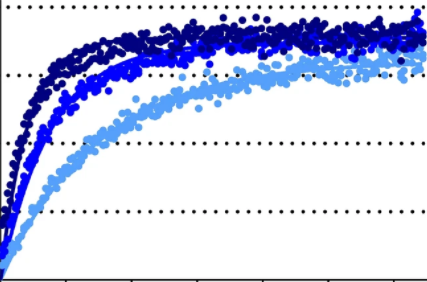
Affinity determination
Step 3
TIRF microscopy imaging and data analysis
Step 4Delivery
Spectral properties of fluorescent ligands
Kinetic and competitive binding of fluorescent ligands
Images of TIRF microscopic imaging
Assessing the effect of fluorophores on the functional activity of ligands
Other data that you need
Our Advantages
- High specificity detection at very low concentrations
- High spatial and temporal resolution
- High affinity and excellent signal-to-noise ratio
- Experienced scientists provide experimental consultation
- Reasonable price and short turnaround time
CD BioSciences has a professional team and advanced imaging equipment. The entire process of dopamine receptors imaging analysis is operated by experienced technicians to ensure the accuracy of the experiment. If you have any needs, please feel free to contact us.
- Allikalt A, Purkayastha N, Flad K, et al. Fluorescent ligands for dopamine D2/D3 receptors[J]. Scientific Reports, 2020, 10(1): 1-13.
- Zhuang Y, Xu P, Mao C, et al. Structural insights into the human D1 and D2 dopamine receptor signaling complexes[J]. Cell, 2021, 184(4): 931-942. e18.
*If your organization requires the signing of a confidentiality agreement, please contact us by email.
Please note: Our services can only be used for research purposes. Do not use in diagnostic or therapeutic procedures!